Electron Microscopy Laboratory
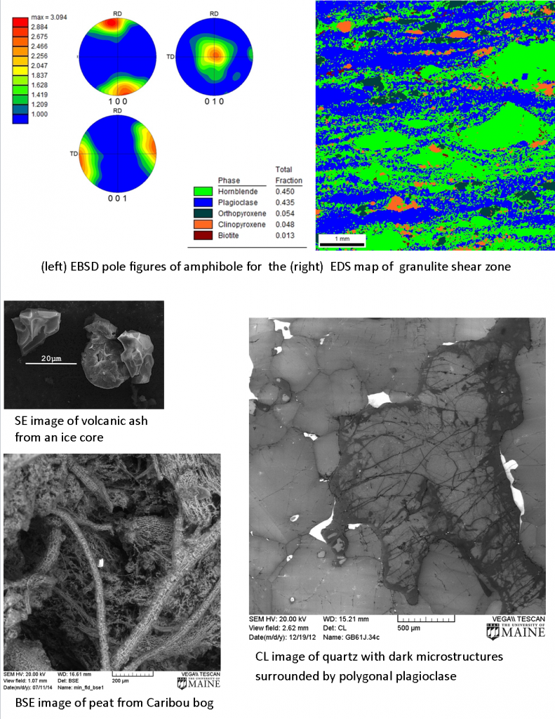
Environmental scanning electon microscope with energy-dispersive elemental analysis, cathodoluminescence imaging, and electron backscatter diffraction capabilities
Contacts: Dr. Chris Gerbi, Dr. Marty Yates, Dr. Won Joon Song
An NSF Major Research Instrumentation grant to Gerbi, Johnson, Grew, Koons, Belknap, Kreutz, Lux, and Yates allowed for the purchase of the equipment in the new laboratory, established in Spring 2009. The lab consists of the following major equipment:
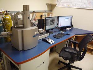
- Tescan Vega II XMU tungsten filament scanning electron microscope
- high vacuum low vacuum (pressures from 5-2000Pa) modes
- backscatter, secondary electron, and low vacuum secondary electron detectors, panchromatic cathodoluminescence detector
- resolution, depth, and wide-field imaging modes
- EDAX Pegasus system
- energy dispersive x-ray spectrometry (EDS) with an Apollo40 SDD
- electron backscatter diffraction (EBSD) with the Digiview IV camera
- Cryo-stage
- maintain temperatures -100C to -120C and high or low vacuum modes
- glove box for transfer of ice samples
- Rotary and vibratory polishing equipment