Electron Microscopy Laboratory
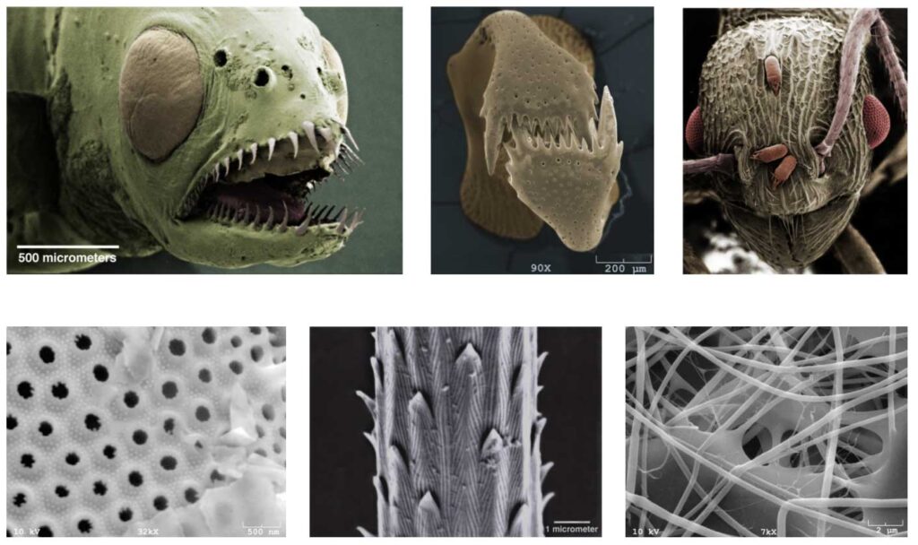
Microscopy provides images of the very small, down to the nanometer scale, and allows analysis of molecular and elemental composition. The EML (Electron Microscopy Laboratory) offers the tools and technical expertise for research and training in microscopy with both light and electron microscopes. We maintain and operate two scanning electron microscopes, a confocal laser scanning microscope, and the ancillary equipment needed for the preparation of specimens for EM and other microscopy. Our multiple light microscopes encompass optics for bright-field, phase-contrast, Nomarski differential-interference, polarizing, and fluorescence imaging.
Users of the EML can opt for full technical services in microscopy so that they are provided with whatever micrographs and analyses are needed for given specimens, or users can work independently on the EML’s microscopes and specimen-preparation equipment following appropriate training by the EML’s staff. Technical service is available for any microscopy-related tasks, such as specimen preparation, microscope operation, and photography.
Individual tutorials on the operation of the EML’s equipment can be arranged.
For more information contact:
Dr. Emma Perry, Electron Microscopy Lab Manager
11 Murray Hall, University of Maine, Orono
207.581.2927
emma.perry@maine.edu.
University of Maine System
Microcredential Pathway for Scanning Electron Microscopy
Electron Microscopy Lab services and rates
Click the microscope icon to:
- See a complete listing of EML equipment and service rates.
- Schedule your next service request.
- Manage your iLab account.
Technical Services
- Microscope operation—full operation of all electron and light microscopes.
- Specimen preservation by chemical and microwave-enhanced fixation.
- Specimen embedment in media suitable for EM or light microscopy.
- Critical-point-drying of specimens for SEM.
- Metal coating of specimens by evaporative or sputter technology.
- Consulting on techniques of microscopy.
Scanning Electron Microscope (SEM)
Scanning Electron Microscope (SEM)for imaging of bulk specimens.
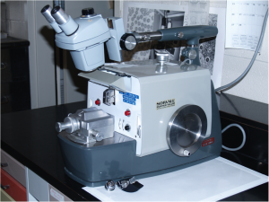
AMRay 1820 Scanning Electron Microscope
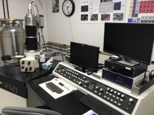
This SEM has a resolution of 5 nm, a magnification range of 20 – 150,000X and an accelerating voltage range of 100 V to 30 kV. Its specimen stage is a eucentric goniometer, meaning that the specimen can be tilted and rotated about the viewing axis. Dynamic focusing accommodates imaging flat specimens. Image capture is fully digital using an iXRF digital-capture system. The microscope is fitted with a high-performance Robinson backscatter detector, and an iXRF EDS system for elemental identification and mapping, in addition to the standard secondary electron detector.
Zeiss NVision 40 FIB/Scanning Electron Microscope
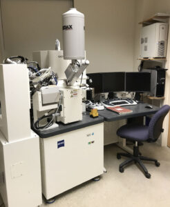
The SEM has a resolution of 1.2 nm, a magnification range of 20X – 600,000X and an accelerating voltage range of 100V to 30KV. It has a six axis eucentric goniometer stage, and multiple detectors including secondary electron, backscatter electron and inlens secondary electron. The microscope is equipped with an EDAX EDS system for elemental identification and mapping.
The FIB has a SIINT zeta Ion Column with ion beam resolution of 4 nm at 30KV, an accelerating voltage range of 3KV to 30KV, and a four channel Gas Injection System.
The NVision 40 is also equipped with;
- Bal-Tec VCT 100 Vacuum-Cryo-Transfer System, with associated cryo-preparation/loading stage, and Bal-Tec SCD 500 sputter coater and QSG 100 Thickness Monitor
- Kleindiek Micromanipulator System
- Raith ELPHY E-beam Lithography System
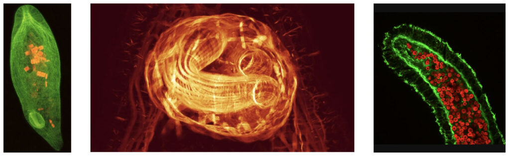
Confocal Laser Scanning Microscopy (CLSM)
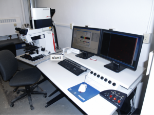
Confocal Laser Scanning Microscopy (CLSM) for optically sectioning thick fluorescent specimens.
Leica TCS SP2 Confocal Microscope — equipped with three lasers (Ar, green HeNE, and red HeNe) with available wavelengths of 457-477 nm, 488 nm, 514 nm, 543 nm, and 633 nm.
Microtomes/Ultramicrotomes
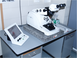
Microtomes and ultramicrotomes — for cutting specimens for both light and electron microscopy. The EML has a suite of four ultramicrotomes—a Leica EM UC6 and three Sorval MT2B ultramicrotomes—for cutting thin sections (40 to 90 nm) for electron microscopy. Additionally, the EML maintains a Sorval JB4 equipped with a Butler trough for serially sectioning resin-embedded specimens for light microscopy (at thicknesses from ½ to 2 μm). All of the microtomes can used with either glass or diamond knives.
Light Microscopes
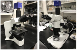
Both compound and dissecting microscopes are available. An Olympus BX41 compound microscope is fitted for standard bright-field, phase-contrast and dark-field imaging. An Olympus SZX16 dissecting microscope is fitted for reflected light, as well as bright-field, oblique, and dark-field illumination from its integral LED stand. Both microscopes are fitted for digital micrography using Zeiss Axiocams.
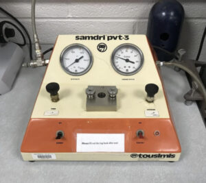
Specimen Drying
Critical–point dryer — for drying SEM specimens without subjecting them to the distorting forces of surface tension.
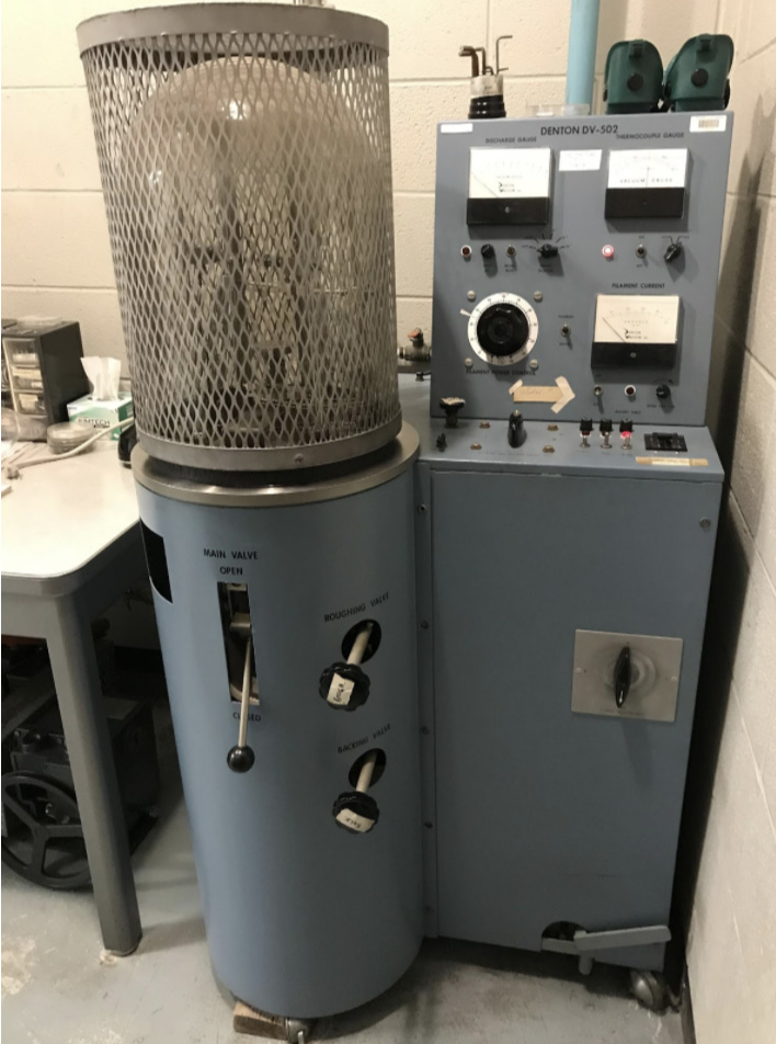
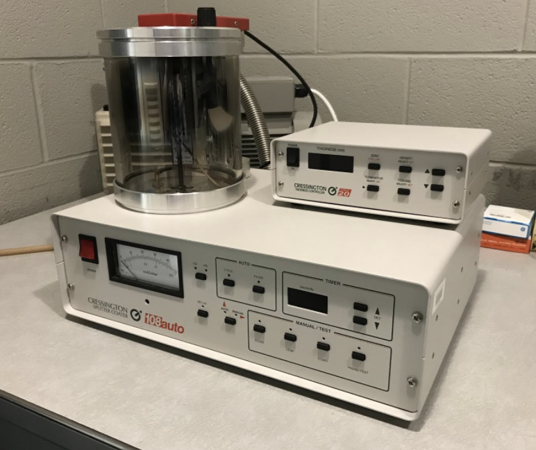
Metal Coating
Metal coating by sputter-coating and vacuum evaporating such metals as gold, gold-palladium, platinum, chromium, aluminum, and carbon. Sputter-coating is typically used for the preparation of SEM specimens, while vacuum evaporation is used for shadowing and producing conductive thin-film supports for EM.