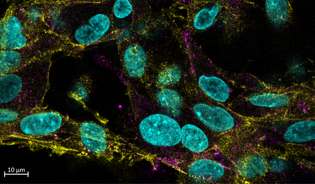
 Normal human astrocytes (NHAs) infected with Alexa Fluor 647-conjugated JC polyomavirus (magenta). Cells were fixed at two hours post infection and stained for pan-Cadherin (yellow) and DAPI (cyan; Sophie Craig, Maginnis Lab).
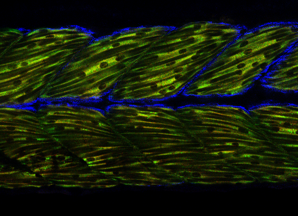 Labeling for mRNA in zebrafish showing overlap of two fast-twitch muscle markers (red/green) adjacent to a slow-twitch marker (blue; Jared Talbot, Talbot Lab)
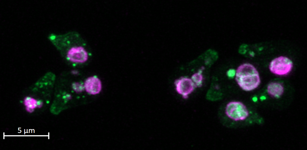 Saccharomyces cerevisiae forming mating projections toward high doses of mating pheromone. The pheromone receptor Ste2 (green) localized at these projections are internalized and traffick from the plasma membrane to the vacuole/lysosome (magenta) following their activation (Nick Leclerc, Kelley Lab).
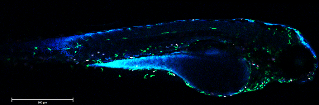 Systematic Influenza A Virus infection and immune cells in zebrafish. 15 hours post infection in zebrafish Tg(mpeg:eGFP;lyz:dsRed) at 3 days post fertilization, Tg(mpeg:eGFP;lyz:dsRed) showing macrophages (green), neutrophils (red), and cyan Color-flu Tg(eCFP-PR8) at 10x resolution (Brandy Soos, King Lab).
 RBL2H3 mast cells stained with Hoechst 33342 (nucleus; blue) and Mitotracker Red FM (mitochondria; magenta) (Esther Biro, Sydni Plummer, and Dylan Wagner, Gosse Lab)
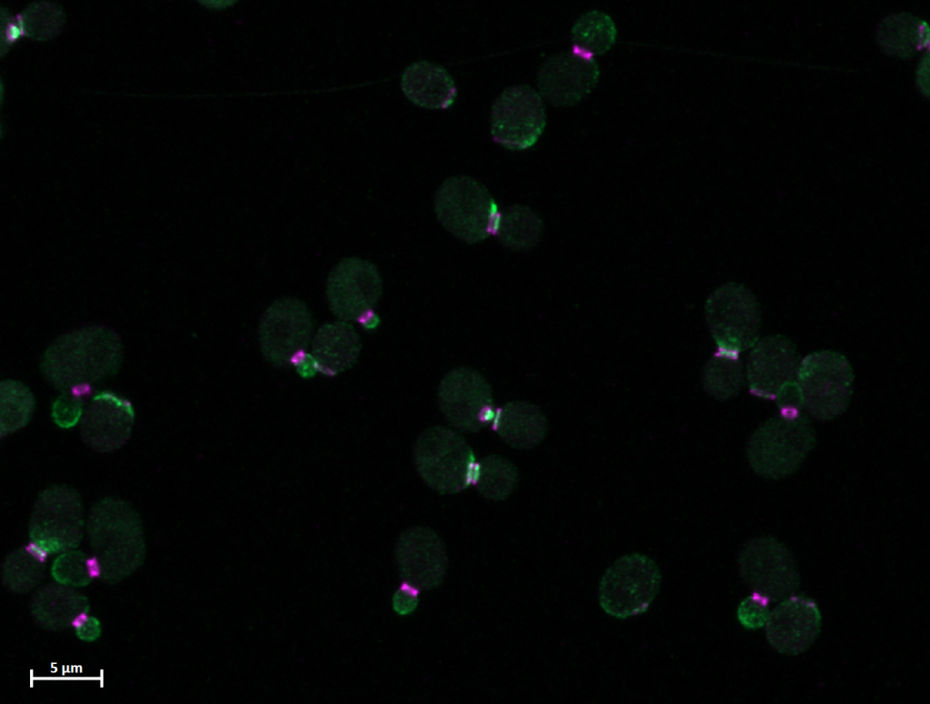 Fluorescence imaging of budding yeast cells (Saccharomyces cerevisiae) using the ZEISS LSM 980 Airyscan. Septin subunit Cdc3 is tagged with mCherry, shown in magenta, while the polar cap protein Bem1 is tagged with GFP, shown in green (Sudati Shrestha, Kelley Lab)
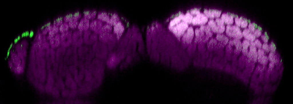 Zebrafish muscle labeled for slow-twitch fibers (green) and fast-twitch fibers (magenta), shown in cross-sectional view (Jared Talbot, Talbot Lab)
 Tracking zebrafish innate immune response to Candida albicans injection into the hindbrain ventricle. Initial C. albicans inoculum injected shown in blue, total C. albicans burden is shown in red, macrophages in green, neutrophils in cyan. Images taken at 24 hours post injection (Nnamdi Baker, Wheeler Lab).
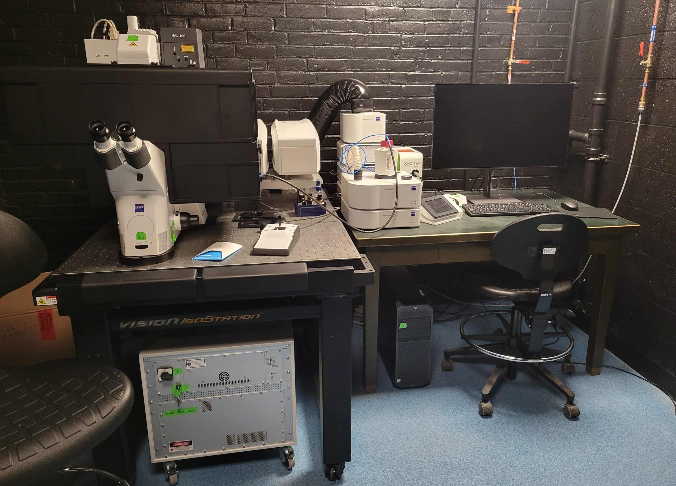
Zeiss LSM980 – Airyscan 2
The inverted laser scanning confocal microscope is ideal for diverse range of applications for confocal and super resolution imaging including multi-color experiments, live sample imaging, protein colocalization, tiling of large samples, Z-stack 3D/4D imaging, and weak label detection. The microscope is equipped with environmental control (temperature, humidity and CO2) and can image live BSL-2 samples. The motorized XY stage can hold samples in slides, 35 mm dishes, chambered slides, and well-plates.
Airyscan 2
• Super resolution with high light efficiency
• Multiplex mode collects image up to 10 times faster
Lasers – all diode lasers
• 405 nm
• 445 nm
• 488 nm
• 561 nm
• 639 nm
Objectives
• EC Plan-Neofluar 10×/0.3 air
• Objective PApo 20×/0.8 high resolution air
• Long working distance 40×/1.1 water
• Objective C PApo 63×/1.4 oil
Detectors
• 2 photomultiplier tubes (PMTs)
• High sensitivity GaAsP detector
• Airyscan detector with 4Y and 8Y fast mode
• Transmitted light T-PMT detector
Software: Zen 3.8 for microscope control, image acquisition and processing
Acknowledging miac:
the NIH requires that all publications containing data collected on the microscope, including press releases, acknowledge NIH grant support. Please use the following format:
The authors would like to acknowledge the Microscopy and Image Analysis Core (MIAC) facility (RRID: RRID:SCR_025784) for use of Zeiss LSM980 Confocal Microscope/Imaris software and staff XXX for helpful training/discussion/collecting the data shown in Figure Y. The MIAC facility was supported by Award number 1P20GM144265 from the National Institutes of Health.