Scott Collins
 Professor of Chemistry; Cooperating Professor of Electrical and Computer Engineering, Prof. Graduate School for Biological Science, co-Director of the Institute for Molecular Biophysics (IMB), member of the Laboratory for Surface Science (LASST); Ph.D. Brigham Young University, 1980
E-mail: scott.collins@maine.edu
Professor Collins did a postdoctoral year in the Department of BioEngineering, University of Utah where he pioneered the emerging field of micro chemical sensors. In 1981 he joined the research staff at Abbott Corporate Laboratories in their diagnostics division developing micro sensor based clinical assays. From 1984-86 Prof. Collins visited the Swiss Centre for Microelectroniques in Neuchatel, Suisse to transfer microsensor technology to the Swiss national microfabrication effort.
In 1986 Prof. Collins returned to the US to start an international consulting company in chemical sensors and microfabrication, and he served as CEO until 2002. In 1992 he accepted an Adjunct Associate Professor position at the University of California, Davis, Department of Electrical and Computer Engineering where he established the MicroInstruments and Systems Laboratory and served as co-Director until 2003. In 2002 Prof. Collins transferred to the University of Maine
Professor Collins’ research interests include the design, fabrication, and development of MicroInstruments, MicroSystems, and NanoDevices across all disciplines; electrochemical characterization of electronic materials; and fractal phase transitions. Below are listed some specific research interests.
| Optical/MicroRobotic Systems |
|
Optomechanical Components. Below: Shown are xyz positioning microstages for optical beam steering and delivery. |
Fourier Transform Spectrometers. Below: Shown is the optical table of a FTIR developed for high altitude atmospheric and space habitat analysis.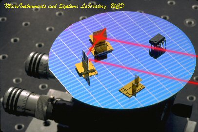 |
Linear Translational Stages. Below: Electrostatic and electromagnetic MEMS translational microstages developed for autofocusing in digital cameras. Stages range in size from less than 1 cm to 8 cm travel distance.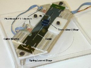 |
|
| MicroNMR Spectroscopy and Imaging |
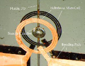 |
Helmholtz Micro Coils. Left: Single three-turn Helmholtz
RF microcoil with nanoliter sample chamber and microfluidic delivery
for high resolution NMR spectroscopy of rare or limited biosamples.
|
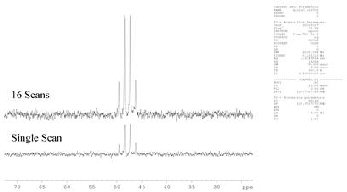
| NMR Spectrum of Acetic Acid. Right: Spectrum of acetic acid showing excellent resolution and S/N even at nanoliter volumes. |
 |
| High-Speed Nanopore Gene Sequencing |
|
Nanopore Gene Sequencing Left: Transition of biopolymer (RNA, DNA, or Protein) modulates tunneling current between electrodes at the pore periphery according to the chemical electron density of the polymer substituent |
| Micrograph of Nanopore Device Right: Metal electrodes span an approximately 10nm nanopore fabricated in a 20nm silicon nitride membrane. Nanopores can be fabricated from 0.7nm to 50nm |
 |
| Aerodynamics and Aerospace Applications |
 |
“Gurney” Microtabs for Airfoils Left: Gurney microtabs
(named after the first Indy driver to use them) on an airfoil.
Actuating the tabs into the airflow increases lift by 50% while
increasing drag by 10%. The micro tabs are used to control and
stabilize aircraft (smart skin). |
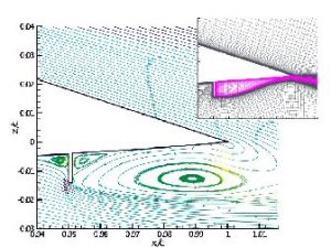
| Recirculation Airflow for Microtabs Right: Computer simulation of recirculation flow at microtabs with a 1% extension at 5% of chord. |
 |
 |
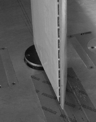
Airfoil with MicroTabs Left: Airfoil in wind tunnel during testing. Microtabs are shown extended a tail end of airfoil. |
| MicroInstruments for Cellular Characterizations |
Cellular Culturing and Testing Left: A micro instrument developed for the testing of endothelial cells for research into arterial sclerosis. MicroInstrument contains multiple microfluidic channels complete with external I/O for controlled perfusion and introduction of hydrodynamic stress. Cells can be probed either optically, with conventional microelectrodes from the top through a
membrane, or with an array of integrated electrodes integrated into the substrate. The instrument is completely modular and can be reversibly disassembled/assembled for sterilization and reuse. |
 |
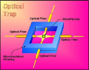 |
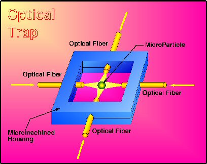
Cytology Micro Workstation Left: An optical trap is the heart of a cellular workstation. Bioparticles are captured with a 4 beam micro optical trap and subsequently analyzed, altered, and/or sorted individually using a suit of optical, mechanical, or acoustic micro instruments appended to the trap. |
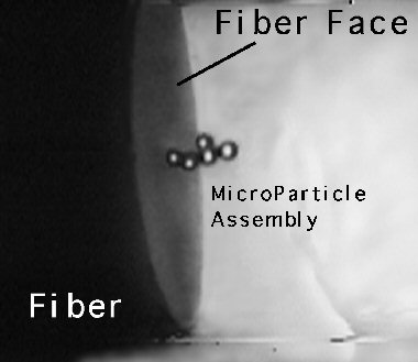
| Cellular Assembly Line Right: The micro optical trap is used to select and assemble individual cells in a specific order at the face of a fiber. The cells are then fused together either chemically or with a laser scalpel. The cells form a cellular chemical assembly line for the biosynthesis of complex molecules or drugs |
 |